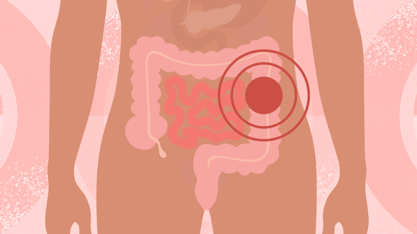
A colonoscopy is the only way to definitively diagnose UC and its subtypes. This procedure is performed under general anesthesia and involves inserting a colonoscope (a long, lighted tube with an attached camera) into your rectum and then into your colon.
A colonoscopy can help doctors see the amount of ulceration and bleeding that has occurred in the mucosa (the lining of the colon).
Lab Tests
Lab tests can include blood work to check for nutrients, antibodies, or signs of infection.
They may also use the stool sample to rule out other possible conditions.
X-rays
Sigmoidoscopy
A flexible sigmoidoscopy is similar to a colonoscopy, but it is less intrusive. This procedure uses a flexible tube with a light to view just the lower portion of the colon, which consists of the two areas leading up to your rectum: the descending colon and the sigmoid colon. A sigmoidoscopy is typically used to evaluate the severity of UC after it’s been diagnosed.
Chromoendoscopy
A chromoendoscopy is a type of colonoscopy that involves injecting a blue dye into the colon. The blue dye helps to highlight possible precancerous lesions and polyps in the colon.
Your doctor may then take a small sample of colon tissue (biopsy) or remove any polyps they find.
Biopsy
If your doctor removes a small tissue sample or a polyp from your colon, they will send it to a lab for examination. The lab can help screen for worsening UC as well as colorectal cancer.
CT Scan
Read the full article here


